Leg Bones Diagram Labeled - Leg Anatomy - Your leg bones are very large and strong to help support the weight of your body.
Leg Bones Diagram Labeled - Leg Anatomy - Your leg bones are very large and strong to help support the weight of your body.. Most bones (particularly the long bones of the arms and legs — which make up the appendicular skeleton) have a hard outer shell known as cortical bone. Labeled human leg bones created for use in leg bone. Its lower end helps create the knee joint. Which of the labeled structures in the diagram is formed as the result of cartilage being replaced by bone after the cessation of bone growth? Human skeletal diagram labeled bones college ruled composition notebook:
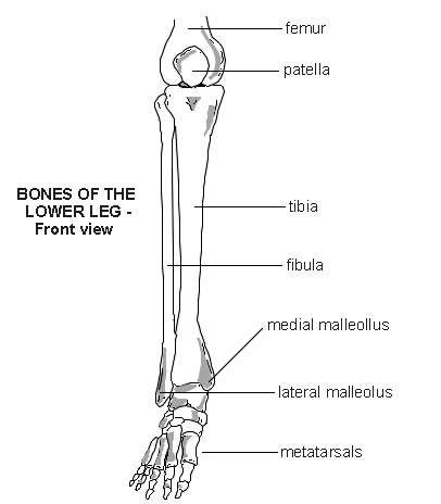
Human skeletal diagram labeled bones college ruled composition notebook: Looking for bone diagram barca fontanacountryinn com? The foot bones shown in this diagram are the talus, navicular, cuneiform, cuboid, metatarsals and calcaneus. Most bones (particularly the long bones of the arms and legs — which make up the appendicular skeleton) have a hard outer shell known as cortical bone. Here's a diagram with the tibia bone labelled, as well as the fibula, showcasing all their surface landmarks.
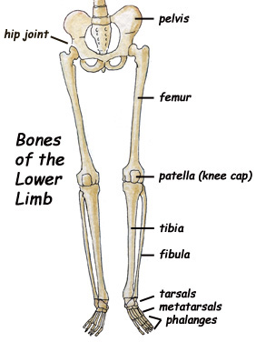
leg muscles diagram - Free Large Images from www.free-largeimages.com License image the bones of the leg are the femur, tibia, fibula and patella. The two legs are attached to hip bone of the skeleton by ball and socket joints. This image is an edited version of this image that was created by user:ladyofhats (mariana ruiz villarreal). The knee joint is the largest joint in the body and is primarily a hinge joint. Start studying labelling leg bones. Which of the labeled structures in the diagram is formed as the result of cartilage being replaced by bone after the cessation of bone growth? *free* shipping on qualifying offers. This framework consists of many individual bones and cartilages.
Frontal skeleton orthopedic anatomy system publishing, castlecomer on amazon.com.
We also discuss what are osteons, what are canaliculi, what are. This image is an edited version of this image that was created by user:ladyofhats (mariana ruiz villarreal). *free* shipping on qualifying offers. Frontal skeleton orthopedic anatomy system publishing, castlecomer on amazon.com. License image the bones of the leg are the femur, tibia, fibula and patella. Labeled medical scheme with humerus, muscle, radius and ulna isolated closeup. Diagram of leg bones, find out more about diagram of leg bones. Study guide for students and teachers. There are 4 intermediate phalanges, one on each finger, except the big toe. The bone that goes from your pelvis to your knee is called the femur (say: Which of the labeled structures in the diagram is formed as the result of cartilage being replaced by bone after the cessation of bone growth? The bones of the leg are the femur, tibia, fibula and patella. This framework consists of many individual bones and cartilages.
Virtual bone labwe need our bones to walk, run, jump and move, but this is not all they do. The bones of your leg have roughened patches on their surfaces where muscles are attached. Bone diagram barca fontanacountryinn com. The two legs are attached to hip bone of the skeleton by ball and socket joints. This image is an edited version of this image that was created by user:ladyofhats (mariana ruiz villarreal).
Skeletal Series Part 10: The Human Leg | These Bones Of Mine from thesebonesofmine.files.wordpress.com Bones are very busy even when you are sleeping at night. Frontal skeleton orthopedic anatomy system publishing, castlecomer on amazon.com. Learn vocabulary, terms and more with flashcards, games and other study tools. Human skeletal diagram labeled bones college ruled composition notebook: This framework consists of many individual bones and cartilages. Which of the labeled structures in the diagram is formed as the result of cartilage being replaced by bone after the cessation of bone growth? Study guide for students and teachers. Looking for bone diagram barca fontanacountryinn com?
The femur, or thighbone, is the longest and largest bone in the human body.
The two legs are attached to hip bone of the skeleton by ball and socket joints. Its lower end helps create the knee joint. Skull, clavicle, mandible, scapula, thorax, sternum, humerus, ulna, radius, carpus, phalanges (fingers), metacarpus, spine, pelvis, sacrum, femur, tibia, fibula, tarsus. This image is an edited version of this image that was created by user:ladyofhats (mariana ruiz villarreal). Which of the labeled structures in the diagram is formed as the result of cartilage being replaced by bone after the cessation of bone growth? The bones of the leg are the femur, tibia, fibula and patella. Here's a diagram with the tibia bone labelled, as well as the fibula, showcasing all their surface landmarks. Which of the labeled structures in the diagram are fragments of older osteons that have been partially destroyed during bone rebuilding or growth? Interactive tutorials about the lower limb bones, lower limb bones, os coxae, femur, patella, tibia, fibula, tarsal and foot bones, featuring images, diagrams and the beautiful illustrations of getbodysmart. The bones mentioned in each human skeleton chart are: The knee joint is the largest joint in the body and is primarily a hinge joint, although some sliding and rotation occur. Leg femur diagram data wiring diagram today. The foot bones shown in this diagram are the talus, navicular, cuneiform, cuboid, metatarsals and calcaneus.
Labeled human leg bones created for use in leg bone. *free* shipping on qualifying offers. Start studying labelling leg bones. Learn vocabulary, terms and more with flashcards, games and other study tools. Bone diagram barca fontanacountryinn com.
Lower leg - bones | Diagram | Patient from patient.azureedge.net In this video we discuss the structure of bone tissue and the components of bones. Each leg consists of three parts: Skull, clavicle, mandible, scapula, thorax, sternum, humerus, ulna, radius, carpus, phalanges (fingers), metacarpus, spine, pelvis, sacrum, femur, tibia, fibula, tarsus. Most bones (particularly the long bones of the arms and legs — which make up the appendicular skeleton) have a hard outer shell known as cortical bone. The bones of the legs are those that make up the thigh, the lower half of the legs, and the feet. Leg femur diagram data wiring diagram today. *free* shipping on qualifying offers. Labeled human leg bones created for use in leg bone.
The bone that goes from your pelvis to your knee is called the femur (say:
Labeled human leg bones created for use in leg bone. Its lower end helps create the knee joint. Your leg bones are the longest and strongest bones in your body. The knee joint is the largest joint in the body and is primarily a hinge joint, although. This image is an edited version of this image that was created by user:ladyofhats (mariana ruiz villarreal). There are 5 proximal phalanges in each foot (as shown in the diagram above). Leg muscle anatomy (front view. At the microscopic level, this hard outer shell is made up of rod like structures called osteons. Which of the labeled structures in the diagram are fragments of older osteons that have been partially destroyed during bone rebuilding or growth? This framework consists of many individual bones and cartilages. The knee joint is the largest joint in the body and is primarily a hinge joint. Most bones (particularly the long bones of the arms and legs — which make up the appendicular skeleton) have a hard outer shell known as cortical bone. In this video we discuss the structure of bone tissue and the components of bones.
The foot bones shown in this diagram are the talus, navicular, cuneiform, cuboid, metatarsals and calcaneus leg bones diagram. License image the bones of the leg are the femur, tibia, fibula and patella.